Professor Yang
 Know more>>
Professor
Department of Chemical Biology, Xiamen University
Know more>>
Professor
Department of Chemical Biology, Xiamen University
Contact Information
Yang's LaboratoryRoom 532, Lujiaxi Building, College of Chemistry and Chemical Engineering, Xiamen University, Xiamen 361005, China
Ph: +86 (0) 592-218 7601cyyang@xmu.edu.cn
Ruolan Huang's paper accepted by Anal Chem
2023-07-31 20:00:00

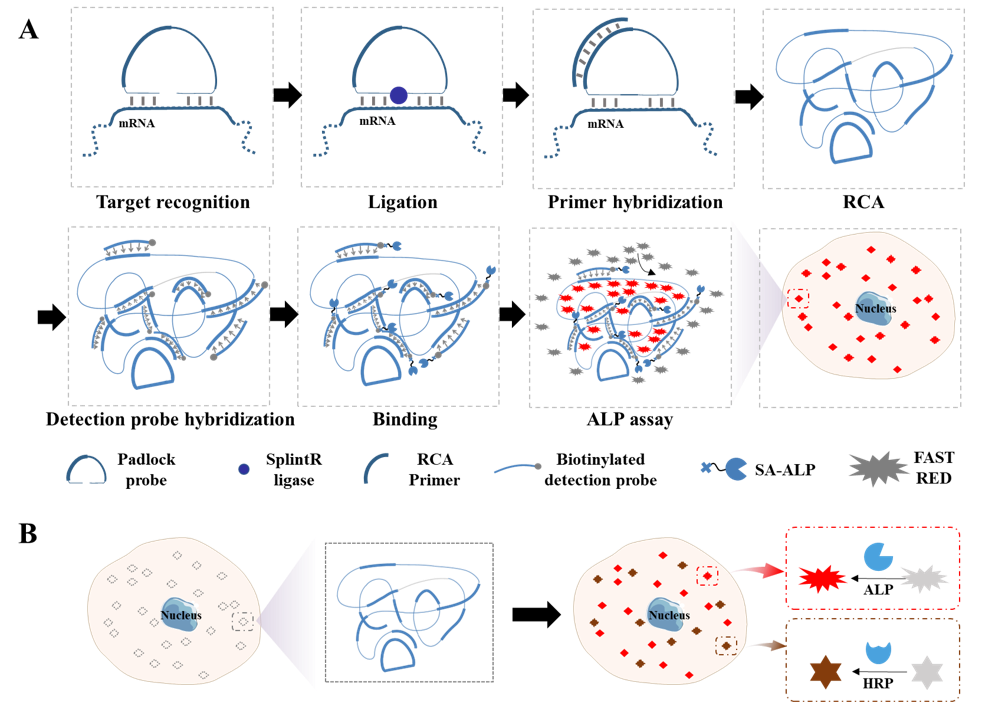
Visualizing spatial patterns of gene expression by optical microscopy at single-molecule resolution represents a long-standing challenge for imaging and molecular engineering technologies. In this study, we developed a method for visualizing mRNA with duplex capability by optical microscopy using rolling circle amplification with streptavidin-modified alkaline phosphatase (SA-ALP) to provide highly selective and sensitive RNA detection. ALP-based RNA detection provides comparable sensitivity and specificity to fluorescence-based in situ assays and similar performance to the current RNAscope technique for single-molecule RNA detection, but with improved ease of operation. This versatile and relatively user-friendly method of single-molecule RNA visualization can also overcome common problems of background interference. Our findings show that the red spots generated by the Fast Red staining in situ are readily quantified by image analysis, even in samples with heavy melanin deposition, supporting the clinical translation of this assay to improve diagnostic assays for recalcitrant tissues. This system was adaptable for duplex assays with multiple probes and multiple stains, which is ALP with horseradish peroxidase to produce red and brown signals to simultaneously visualize two different RNA targets. The duplex assay can be successfully applied to quantify mRNA expression from two genes in situ within single cells and multiple cell types. With the advantages of high sensitivity and low hardware requirements, this versatile and user-friendly method of RNA visualization may enable low-resource institutions to conduct previously inaccessible diagnostic or research questions about the in situ expression and distribution of RNAs at single-molecule resolution.